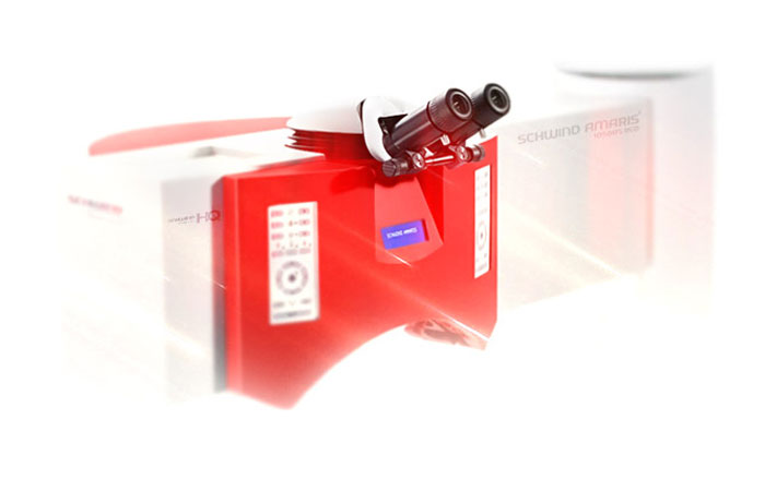
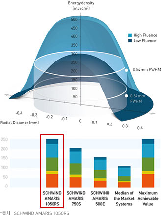
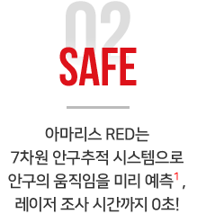
사람마다 각막의 각 지점별로 각막 상피두께가 모두 다릅니다. 본원의 아마리스 레드 1050RS 장비에 탑재된 최신 스마트펄스 알고리즘의 소프트웨어를 통해 수술 장비에 입력한 수술자 고유의 각막 상피두께값 그대로, 1 마이크론 단위의 정밀 수술이 가능합니다. 불필요한 각막절삭과 과ㆍ오교정으로부터 안심하기 위한 진정한 1:1 각막지형 맞춤수술을 만나보세요.
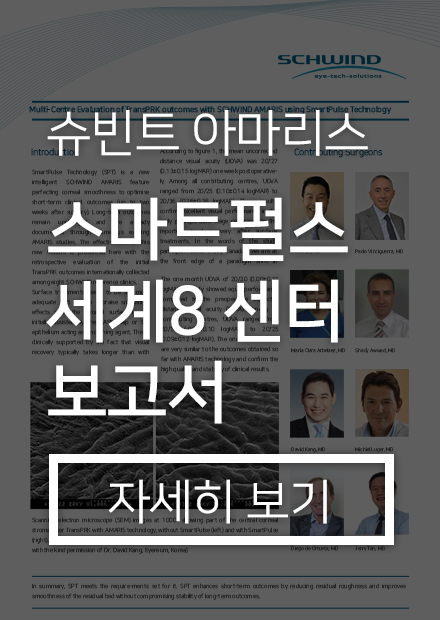
Name
Location
David T Lin
@ Vancouver, Canada
Michiel HA Luger
@ Utrecht, The Nether Lands
Diego de Ortueta
@ Recklinghausen, Germany
Shady T Awwad
@ Beirut, Lebanon
Paolo Vinciguerra
@ Milano, Italy
Jerry Tan
@ Singapore
Maria C Arbelaez
@ Muscat, Oman
David Sung-Yong Kang
@ Seoul, Korea
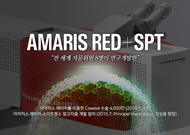
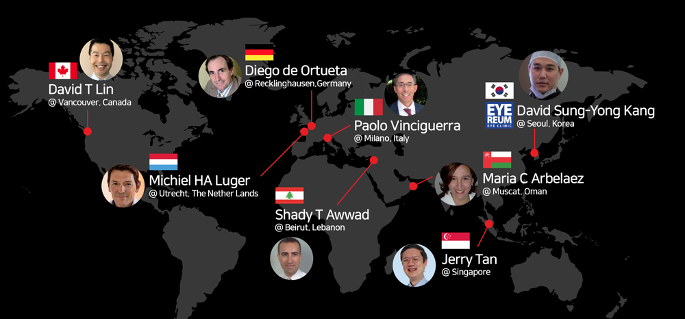
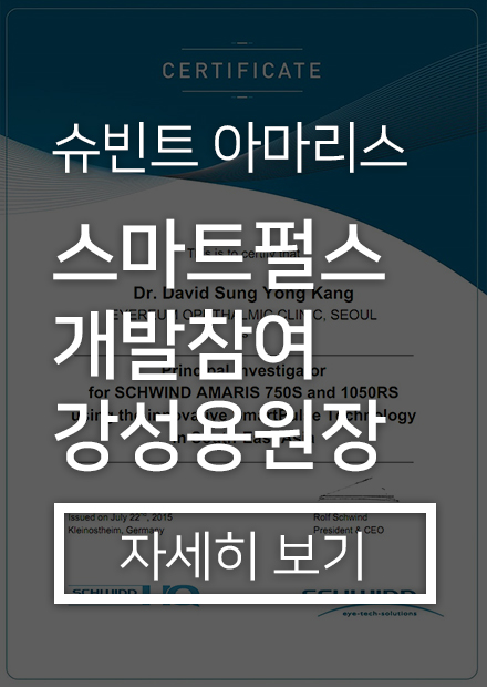
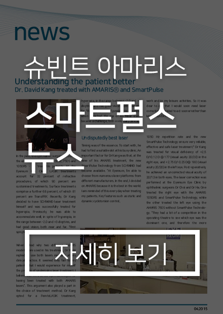
아마리스레이저의 3D 알고리즘, 스마트펄스(AMARIS Smart Pulse) 개발 프로젝트를 위해 한국을 비롯하여 이탈리아, 독일, 네덜란드, 캐나다, 싱가포르 등 8개국에 아마리스 스마트펄스(SPT) 연구를 위한 안과 의사들이 참여했습니다. 당시 한국에서는 유일하게 아이리움안과 강성용 원장이 본 프로젝트에 참여했습니다. 슈빈트 아마리스레이저의 스마트펄스 알고리즘은 현재 Smartsurface 라는 이름으로 제공되고 있으며 개발 후 The Ophthalmologist 에서 Innovation Award 2015에 소개하기도 하였습니다.
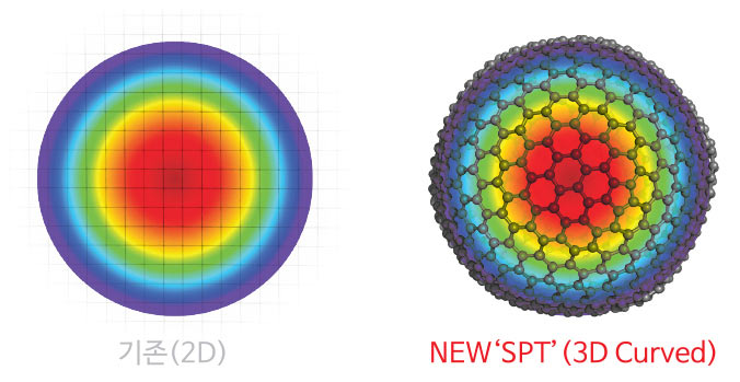
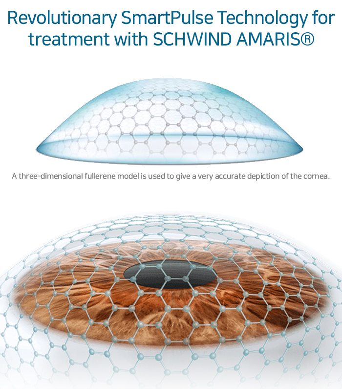
스마트펄스 테크놀로지(SPT)는 현존하는 레이저 알고리즘 중 실제 우리 눈의 각막 곡률을 가장 사실적으로 구현한 레이저 조사 방식입니다. 사람의 눈은 축구공처럼 구 모양이기 때문에 각막의 실제 곡률을 고려한 입체적인 레이저 수술기법이 필요했습니다. 기존 방식은 편평한 2차원 좌표 위에 레이저를 조사할 지점의 결정하지만, 새로운 알고리즘, 스마트 펄스 테크놀로지(SPT)는 입체적인 탄소분자모형 구조(Fullerene Structure)를 기반으로 각막 곡률에 맞는 3차원 좌표(x, y, z) 값을 정해 구 모양의 각막에 입체적으로 레이저를 조사합니다.
수술 후 각막 표면이 부드러워야 하는 이유
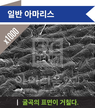
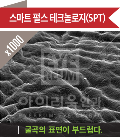
안경렌즈 표면이 매끄러우면 더 선명하듯이 우리 눈의 안경 렌즈와 같은 각막 표면이 매끄러울수록 시력의 질은 당연히 높아집니다. 기존 레이저 시력교정수술 후에는 각막 표면이 거칠 어진 상태로 남지만 아마리스 레이저의 스마트펄스 기술은 각막 표면을 가장 부드럽게 만들어 수술 직후부터 선명한 시력을 찾을 수 있습니다.
라섹과 같은 표면절제술에서 스마트펄스의 효과가 더욱 분명히 나타납니다. 실제 라섹 수술 환자의 각막 표면의 거친 정도를 특정한 수치를 비교해보면, 기존 레이저로 수술했을 때는 평균 749nm, 스마트펄스로 수술한 경우에는 평균 272nm로 기존의 1/3 수준으로 향상된 결과를 보였습니다. (2014. 9 ESCRS, Paolo, Italy)
일반 아마리스
x1000
굴곡의 표면이 거칠다.
스마트펄스테크놀로지(SPT)
x1000
EYEREUM 아이리움안과 EYEREUM EYE CLINIC
굴곡의 표면이 부드럽다.
▲ 타 레이저와의 수술 후 각막 표면 비교 - 아이리움안과/연세대 세브란스 공동, ⓒEYEREUM, All rights reserved.
Smart Pulse Technology
스마트 펄스 테크놀로지(SPT ; Smart Pulse Tech
nology)는 수술 후 잔여 각막 층을 가장 부드럽게 하여 단기간 내 시력을 향상시키는 3차원 입체 레이저 알고리즘 입니다. 각막의 실제 곡률에 가장 가깝게 설계해 빠른 시력회복은 물론 시력의 질, 회복기간과 통증, 고위수차 감소에 획기적인 기술입니다.
Revolutionary SmartPulse Technology for treatment with SCHWIND AMARIS®
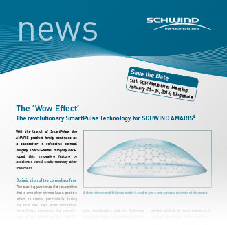
A three-dimensional fullerene model is used to give a very accurate depiction of the cornea.
전 세계 8개 스마트펄스 공식 연구센터의 임상결과 SPT는 수술 후 각막 표면을 부드럽게 하여 수술 후 시력의 질을 혁신적으로 향상시켰고 회복기간과 통증감소, 수술 후 빛 번짐 예방 및 절편 이탈 예방에 효과적입니다.
SCHWIND eye-tech-solutions news
Save the Date
14th SCHWIND User Meeting
January 21-24, 2014, Singapore
The 'Wow Effect' The revolutionary SmartPulse Technology for SCHWIND AMARIS®
With the launch of SmartPulse, the AMARIS product family continues as a pacesetter in refractive corneal surgery. The SCHWIND company developed this innovative feature to accelerate visual acuity recovery after treatment.
A three-dimensional fullerene model is used to give a very accurate depiction of the cornea.
Optimization of the corneal surface
The starting point was the recognition that a smoother cornea has a positive effect on vision, particularly during the first few days after treatment. SmartPulse optimizes the smoothness of the corneal surface...
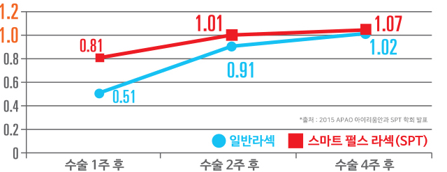
스마트펄스(SPT) 공식센터 아이리움안과가 수술 전 평균 -7디옵터 이상이었던 환자들에게 스마트 펄스(SPT)로 라섹(LASEK) 수술을 시행한 결과 수술 1주 내 시력 회복 속도가 기존 방식보다 1.6배 더 빨랐고 수술 2주 후 평균시력 1.0을 달성했습니다.
아마리스레드 스마트펄스 테크놀로지(SPT) 효과
각막 절삭면을 매끈하게
울퉁불퉁하고 불규칙한 각막 절삭면을 부드럽고 매끈하게 교정
빠른 회복
각막 표면이 매끄러워 각막 상피 재생을 빠르게 하고 통증을 줄임
라섹 수술 2주 후 평균 1.0 달성
수술 후 매끈한 각막절제면, 빠른 회복을 가능케 하는 SPT 공동개발과 각막지형 맞춤수술
‘코웨이브(커스텀아이즈, CustomEyes)’ 수술 건수 40,000안 돌파(2011~2018)
01아마리스 RED는 디옵터 당 각막 절삭 시간이 1.3초 미만으로 현존하는 엑시머레이저 중 가장 빠른 각막 절삭 속도(1050HZ)(2014년 기준)
02아마리스 RED는 각막 절삭 속도가 빨라지면서 각막 세포의 열 손상을 획기적으로 예방
03아마리스 RED는 현존하는 장비 중 가장 절삭량이 적은 플라잉-스팟 (Flying-Spot) 방식의 레이저(2014년 기준)
04현존하는 장비 중 가장 절삭량이 적은 플라잉-스팟(Flying-Spot) 방식의 레이저(2014년 기준)
05업그레이드 된 동공 중심 포착 기능과 각막 두께 실시간 측정 기능
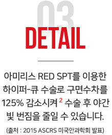
06수술 후 발생할 수 있는 구면수차를 125% 감소시키는 맞춤형 수술 하이퍼-큐 가능
07업그레이드 된 SCC(주시점 보정) 모드를 통한 시력의 질 개선
08수술 중 외부 영향을 받지 않는 자체 제어 시스템 강화
※ 2 출처 : Comparison of Post Operative High Order Aberrations after Trans PRK between Two Types of Ablation Protocols: Aberration Free and Cornea Wavefront-Guided, Using a High Repetition 1050Hz Excimer Laser. 2015, 미국안과학회 ASCRS 발표, 아이리움안과
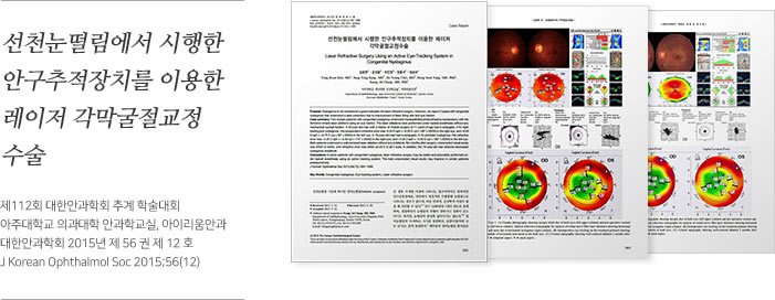
선천눈떨림에서 시행한 안구추적장치를 이용한 레이저 각막굴절교정 수술
학술지 및 발표 정보
제112회 대한안과학회 추계 학술대회
아주대학교 의과대학 안과학교실, 아이리움안과
대한안과학회 2015년 제 56 권 제 12 호
J Korean Ophthalmol Soc 2015;56(12)
Case Report: Laser Refractive Surgery Using an Active Eye-Tracking System in Congenital Nystagmus
Purpose: Nystagmus is not considered a good indication for laser refractive surgery. However, we report 2 cases with congenital nystagmus that underwent a safe procedure due to improvement of laser firing and eye-tracker.
Case summary: Two female patients with congenital nystagmus underwent laser-assisted subepithelial keratectomy with the Schwind Amaris laser platform using an eye-tracker. The laser ablations were performed under topical anesthesia without any mechanical eyeball fixation. A 20-year-old man with a history of mobile surgery at 5 years of age had a conjugate, jerky, right beating jerk nystagmus. The preoperative refractive error was -6.00 D sph = -0.50 D cyl x 180° (0.15) in the right eye, and -6.25 D sph = -0.75 D cyl x 90° (0.05/0.2) in the left eye. A 19-year-old man had a conjugate, horizontal nystagmus. His refractive error was -3.25 D sph = -0.50 cyl x 170° (1.0/0.8) in the right eye, and -3.25 D sph = -0.50 D cyl x 180° (0.9/0.8) in the left eye. Both patients underwent a safe personal laser ablation without any problems. Six months after surgery, uncorrected visual acuity was 20/20 or better, and refractive error was within ±0.25 D in all 4 eyes. In addition, the 19-year-old man showed decreased nystagmus amplitude.
Conclusions: In some patients with congenital nystagmus, laser refractive surgery may be safely and accurately performed under topical anesthesia using an active tracking system. The best uncorrected visual acuity may improve in certain patients postoperatively.
Key Words
Congenital nystagmus, Eye-tracking system, Laser refractive surgery
'안구진탕 환자의 성공적인 시력교정술 사례'
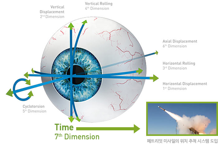
그동안 눈동자의 떨림이 심한 안구진탕 환자의 경우 정확한 위치에 레이저를 조사하는데 어려움이 있어 라식이나 라섹과 같은 시력교정술의 기회조차 얻기 힘든 경우가 많았으나 최신 아마리스 레드 1050RS는 독보적인 7차원 안구추적 시스템과 1050hz의 레이저 조사속도로 추적 즉시 레이저 조사가 가능한 1대 1 시력교정술이 가능해졌습니다.
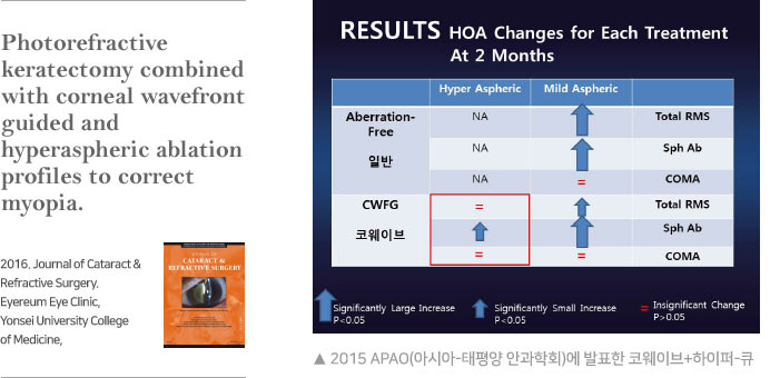
Photorefractive keratectomy combined with corneal wavefront guided and hyperaspheric ablation profiles to correct myopia.
2016, Journal of Cataract & Refractive Surgery. Eyereum Eye Clinic, Yonsei University College of Medicine,
RESULTS HOA Changes for Each Treatment At 2 Months
Treatment
Hyper Aspheric
Mild Aspheric
Aberration Type
Aberration-Free 일반
NA
Significantly Small Increase
Total RMS
Aberration-Free 일반
NA
Significantly Small Increase
Sph Ab
Aberration-Free 일반
NA
Insignificant Change
COMA
CWFG 코웨이브
Insignificant Change
Significantly Small Increase
Total RMS
CWFG 코웨이브
Significantly Small Increase
Significantly Small Increase
Sph Ab
CWFG 코웨이브
Insignificant Change
Insignificant Change
COMA
Legend
Significantly Large Increase P<0.05
Significantly Small Increase P<0.05
= Insignificant Change P>0.05
▲ 2015 APAO(아시아-태평양 안과학회)에 발표한 코웨이브+하이퍼-큐
아마리스 레드 SPT 하이퍼-큐, 구면수차 125% 감소!
최근 SCI(E) 등재 학술지 JCRS에 채택된 코웨이브(커스텀아이즈, CustomEyes) 하이퍼-큐 논문 ‘Photorefractive
keratectomy combined with corneal wavefront–guided and hyperaspheric ablation profiles to correct myopia.’ 라섹 수술 시 절삭량은 보존하면서 빛 번짐은 줄이는 하이퍼-큐 라섹(Hyper-Q)의 연구 내용은 다음과 같습니다.
아마리스 RED SPT를 이용한 하이퍼-큐 라섹과 코웨이브(커스텀아이즈, CustomEyes) 라섹, 그리고 일반 라섹 수술의 2개월 후 결과를 비교하였더니 하이퍼-큐 라섹의 경우에서 구면수차가 125% 감소되는 것을 확인할 수 있었습니다.
아마리스 RED (1050RS) vs 타사 레이저
구분
아마리스 RED (1050RS)
타사 레이저
레이저 빔 종류
Super Gaussian - Flying Spot (only)
3D SmartPulse Technology
7차원 안구 추적 (X, Y, Z, X Rotation,
Y Rotation, Static Cyclo Torsion,
Dynamic Cyclo Torsion, Time)
최대 6차원 (X, Y, Static Cyclo Torsion)
안구 추적 속도
1050 Hz (안구의 움직임과 Laser 절삭
속도보다 더 빠른 인지 속도)
1050 Hz 이하
안구추적과 레이저조사
반응 시간 차
추적 즉시 레이저 조사 (Latency-Free)
3 ms 이상
실시간 각막 측정
Yes (수술 중 각막 절제량 제어 가능)
No
자동 동공 크기조절
Yes (검사와 수술 시, 동공의 크기를
맞춰 최적의 Wavefront 수술 환경
적용)
No
Wavefront 진단 장비
Corneal / Ocular
(전체 수치와 각막 수치를 분리해서
수술할 수 있는 진단 장비)
Topo or Ocular
EYEREUM ONLY
Corneal Biomechanics Exam
원추각막의 가능성을 찾아내는 각막 생체 역학 검사
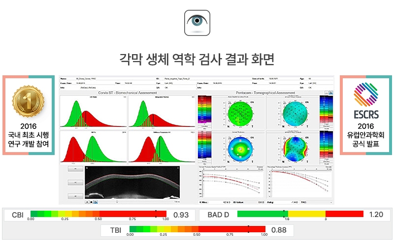
각막 생체 역학 검사 결과 화면
구분
내용
2016 국내 최초 시행 연구 개발 참여
국내 최초 시행 및 연구 개발 참여 성과
ESCRS 2016 유럽안과학회 공식 발표
유럽안과학회 공식 발표 성과
Corvis ST - Biomechanical Assessment / Pentacam - Tomographical Assessment
지표
범위 시각화
수치
CBI
0.00 (Green) to 1.00 (Red) scale
0.93
BAD D
1.6 (Green) to 3 (Red) scale
1.20
TBI
0.00 (Green) to 1.00 (Red) scale
0.88
▲ 각막 생체 역학 검사 결과를 보여주는 평가 그래프
(CBI : Corvis Biomechanical Index / BAD D : Belin / Ambrosio D / TBI : Tomographic Biomechanical Index)
각막 생체 역학 검사는 기존 코르비스ST 각막 강성도 검사와 팬타캠 각막 검사 결과를 비교하여 수술 전 각막의 생체 역학적 상태를 가장 정밀하게 분석해줍니다.
오직 안전을 위한 병원다움
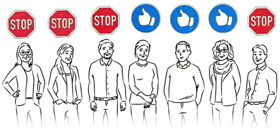
각막 생체 역학 검사는 위험한 수술을 멈추게 합니다.
Person 1
Person 2
Person 3
Person 4
Person 5
Person 6
Person 7
STOP
STOP
STOP
Thumbs Up
Thumbs Up
Thumbs Up
STOP
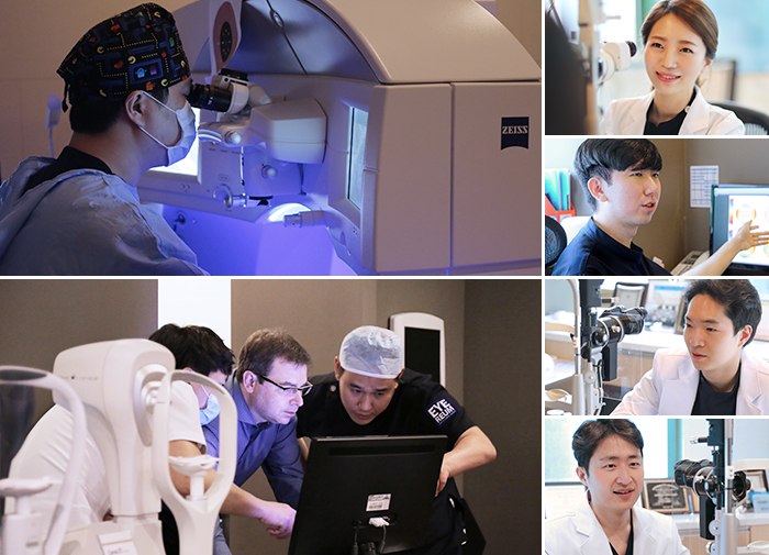
코르비스ST 각막 강성도 검사와 각막 지형도 검사의 결합 분석 프로그램은 아이리움안과의 강성용 원장님과 각막 생체 역학(Corneal Biomechanics) 분야의 권위자 Cynthia Roberts 박사, Renato Ambrosio 박사, Paolo Vinciguerra 교수와 함께 공동개발을 진행했습니다.
신뢰할 수 있는 의료서비스를 위해
SCI 논문으로 과학적 검증을 거칩니다
SCI(Science Citation Index)
과학기술논문 인용색인(SCI)란, 미국 클래리베이트 애널리틱스가 구축한 국제학술논문 데이터베이스로, 매년 학술적 기여도가 높은 학술지를 엄선하고, 학술지에 수록된 논문의 색인 및 인용정보를 데이터베이스화하여 제공하고 있습니다. 더 안전하고 좋은 수술을 만들기 위해, 꾸준한 학술활동과 임상연구에 힘쓰겠습니다.
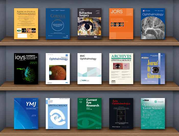
Ophthalmology and Vision Science Journals
Journal Title
Details
American Journal of Ophthalmology
AJO
CORNEA
The Journal of Cornea and External Disease
Journal of Refractive Surgery
Official Journal of the International Society of Refractive Surgery
JCRS
Journal of Cataract & Refractive Surgery
Ophthalmology
Journal of the American Academy of Ophthalmology
iovs
Investigative Ophthalmology & Visual Science
British Journal of Ophthalmology
bjo.bmj.com
BMC Ophthalmology
BioMed Central
ARCHIVES OF OPHTHALMOLOGY
JAMA Network
Journal of Glaucoma
Volume 22, Number 5, June/July 2013
YMJ
YONSEI MEDICAL JOURNAL
JAPANESE JOURNAL OF OPHTHALMOLOGY
Springer
Current Eye Research
Volume 33, Number 11, November 2008
Acta Ophthalmologica
Official Journal of the Nordic and Dutch Ophthalmological Societies
Optometry and Vision Science
Official Publication of the American Academy of Optometry