[소식] 2023 유럽백내장굴절수술학회- 아이리움안과 스마일라식, 노안교정술 강연
아이리움안과의 2023유럽백내장굴절수술학회(ESCRS)
초청 강연 소식을 전해드립니다.

VIENNA 2023
국제적으로 가장 권위있는 안과학회 중 하나인
2023 ESCRS가 지난 9월 8일 -12일
오스트리아 빈에서 진행되었습니다..
이번 학회에서 아이리움안과 의료진들은
스마일라식, 노안교정술의 임상결과와
최신 수술 트렌드를 전했습니다.
41st CONGRESS OF THE ESCRS MESSE WIEN EXHIBITION & CONGRESS CENTRE
VIENNA, AUSTRIA 8-12 SEPTEMBER 2023 Session Rooms: Arena, C2-C5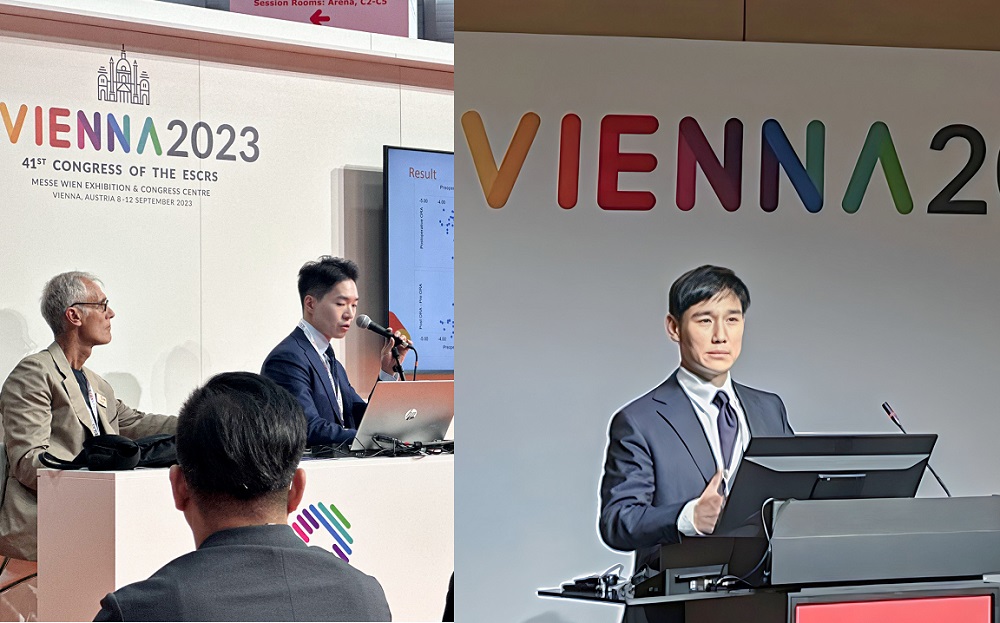
VIENNA 2023
Result
(좌) 아이리움안과 정병훈 원장 / (우) 강성용원장
● What is new with SMILE?
- 스마일 난시교정 해법 '벡터플래닝(Vector Planning)'
스마일수술로 고도난시 교정 시
정확도를 높이고
안구잔여난시(ORA)를 최소화하기 위한
스마일 벡터플래닝 강연
August 2020 Volume 46 Issue 8 Ik-Hee Jun, MD, PhD, David Sung Yong Kang, MD, Samuel Arba-Mosquera, PhD, Noel Alpins, AM, FRANZCO, FRCOphth, FACS, Dan Reinstein, MD, Seong-Ik Jeon, BS, Bong-Kwon Kim, MD, PhD, Kyoung Yul Seo, MD, PhD, Hyung Keun Lee, MD, PhD, Tae-im Kim, MD, PhD J Cataract Refract Surg 2020; 46:1-11 Copyright © 2020 Published by Wolters Kluwer on behalf of ASCRS and ESCRS In laser corneal refractive surgeries for astigmatism correction, 2 treatment strategies are used, based on the manifest refraction or the shape of the cornea. Although the cylinder of the manifest refraction is mainly caused by anterior corneal astigmatism, corneal topographic astigmatism is not always consistent with the refractive cylinder. The difference between corneal astigmatism and refractive cylinder at the corneal plane can be described as the ocular residual astigmatism (ORA), calculated as the vectorial difference. This difference is caused by the posterior corneal surface and crystalline lens, as well as by nonoptical perceptive factors. Previous studies have shown that ORA in the healthy astigmatic eyes that underwent corneal refractive surgery was fairly large, with a mean of 0.73 to 0.81 diopters (D), and even 34% to 60% over 1.0 D. High ORA has been shown to negatively affect the postoperative visual and refractive outcomes after... From the Department of Ophthalmology (Jun, B.-K. Kim, T. Kim), the Institute of Vision Research, Yonsei University College of Medicine, Department of Ophthalmology (Jun, B.-K. Kim, T. Kim), Corneal Dystrophy Research Institute, Yonsei University College of Medicine, Eyereum Eye Clinic (Kang, Jeon), Seoul, South Korea; and Research and Development (Arba-Mosquera), SCHWIND eye-tech-solutions, MS, and Alpins & St. George's Eye Centre (Alpins), Melbourne, Australia; and London Vision Clinic (Reinstein), London, United Kingdom; and Department of Ophthalmology (Reinstein), Columbia University Medical Center, NY, USA; and Centre Hospitalier National d'Ophtalmologie (Reinstein), Paris, France; and Biomedical Science Research Institute (Reinstein), University of Ulster, Coleraine, Northern Ireland. Supported by a grant of the Korea Health Technology R&D Project through the Korea Health Industry Development Institute (KHIDI), funded by the Ministry of Health and Welfare, Republic of Korea (grant number: HI18C1116). The funding organization had no role in the design or conduct of this study. Copyright © 2020 Published by Wolters Kluwer on behalf of ASCRS and ESCRS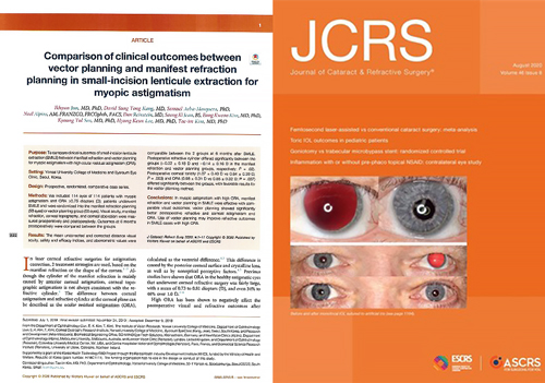
JCRS Journal of Cataract & Refractive Surgery®
ARTICLE
Comparison of clinical outcomes between vector planning and manifest refraction planning in small-incision lenticule extraction for myopic astigmatism
▲ 2020년 8월, SCI 안과학저널 JCRS에 등재된 스마일 벡터플래닝
● 노안치료의 표준, 프레즈비맥스(PresbyMAX)노안교정술
- 근시와 노안을 동시 교정하는
새로운 혼합시력 접근법
프레즈비맥스 노안라식의
강성용 원장 수술 노하우 전수
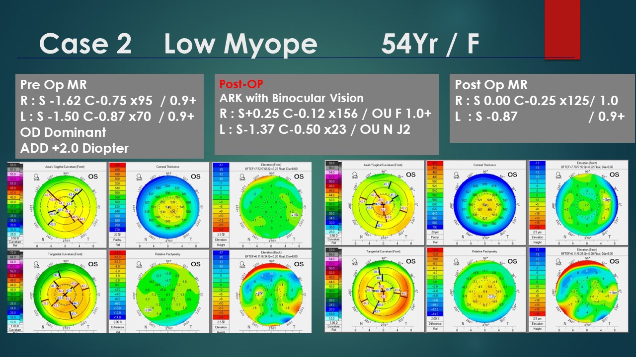
Case 2 Low Myope 54Yr / F
Category Details Pre Op MR R : S -1.62 C-0.75 x95 / 0.9+
L : S -1.50 C-0.87 x70 / 0.9+
OD Dominant
ADD +2.0 Diopter Post-OP ARK with Binocular Vision R : S+0.25 C-0.12 x156 / OU F 1.0+
L : S-1.37 C-0.50 x23 / OU N J2 Post Op MR R : S 0.00 C-0.25 x125/ 1.0
L : S -0.87 / 0.9+ Corneal Topography Analysis - OS
Map Type Description/Parameters Axial / Sagittal Curvature (Front) OS, Curvature Rel Corneal Thickness OS, Pachy Rel Elevation (Front) OS, BFTEF=7.52/7.65 Q=-0.22 Float, Dia=8.00 Tangential Curvature (Front) OS, Curvature Rel Relative Pachymetry OS, Difference Rel Elevation (Back) OS, BFTEF=6.11/6.34 Q=-0.20 Float, Dia=8.00 Corneal Topography Analysis - OS (Post-Op Comparison)
Map Type Description/Parameters Axial / Sagittal Curvature (Front) OS, Curvature Rel Corneal Thickness OS, Pachy Rel Elevation (Front) OS, BFTEF=7.78/7.92 Q=-0.22 Float, Dia=8.00 Tangential Curvature (Front) OS, Curvature Rel Relative Pachymetry OS, Difference Rel Elevation (Back) OS, BFTEF=6.11/6.26 Q=-0.20 Float, Dia=8.00
▲ 프레즈비맥스 노안교정 강연 내용 발췌
아이리움안과는 앞으로도 꾸준한 연구활동으로
신뢰도 높은 의료서비스를 환자분들께 제공하기 위해
노력하겠습니다.