NOTICE & NEWS
EYEREUM OPHTHALMIC CLINIC
ESCRS 37TH CONGRESS OF THE ESCRS PARIS
OCULUS - PENTACAM Holladay Report
Lowering laser energy levels can improve surface quality of the lenticule of SMILE.(Dr. David Kang , Jin Young Choi)
J Refract Surg. 2017 Sep 1;33(9):617-624. doi: 10.3928/1081597X-20170620-02.
Effect of Lowering Laser Energy on the Surface Roughness of Human Corneal Lenticules in SMILE.
Yong Woo Ji, MD; Minseo Kim; David Sung Yong Kang, MD; Dan Z. Reinstein, MD, MA(Cantab), FRCSC; Timothy J. Archer, PhD; Jin Young Choi, MD; Eung Kweon Kim, MD, PhD; Hyung Keun Lee, MD, PhD; Hyoung Yul Seo, MD, PhD; Tae-im Kim, MD, PhD PURPOSE: To determine the effect of lowering femtosecond laser energy on the surface quality of the intrastromal interface during small incision lenticule extraction (SMILE). METHODS: Forty age- and diopter-matched female patients (40 eyes) with moderate myopia received SMILE with different energy levels (100 to 150 nJ) and fixed spot separation (4.5 μm). Five human corneal lenticules from each energy group were evaluated by atomic force microscopy and scanning electron microscopy (SEM). Both anterior and posterior surface characteristics of the lenticules were assessed. RESULTS: All measurements of surface roughness were approximately three times higher and in the anterior and posterior surface of the lenticules with the energy level of 150 nJ than with 100 nJ (P < .001). Furthermore, atomic force microscopy analysis found that energy differences of 15 nJ or more made a significant difference in surface roughness at energy levels of 115 nJ or higher. Interestingly, there was no significant difference in all roughness values of both surfaces among the 100, 105, and 110 nJ groups. In addition, all values of surface roughness were significantly positively correlated with laser energy for both anterior and posterior surfaces of the lenticule (P < .001). Consistent with atomic force microscopy results, SEM also showed that the SMILE lenticules in the higher laser energy group had more irregular surfaces. CONCLUSIONS: Lowering laser energy levels can improve surface quality of the lenticule of SMILE. To achieve better visual outcomes with faster recovery after the procedure, it is recommended to reduce the laser energy to less than 115 nJ at a spot separation of 4.5 μm. [J Refract Surg. 2017;33(9):617-624.] Small incision lenticule extraction (SMILE) is the latest refractive surgical technique using only a femtosecond laser, which removes the corneal stromal lenticule through a small corneal incision ranging from 2 to 5 mm.1,2 This procedure could be achieved by using a femtosecond laser platform with higher firing frequency. Recently, the 500-kHz VisuMax (Carl Zeiss Meditec AG, Jena, Germany) platform with a pulse energy of 115 to 170 nJ has been used.1,3,4 Several studies have reported comparable or superior efficacy, predictability, and safety to other refractive surgery techniques, including femtosecond lenticule extraction (FLEx).4-7 However, SMILE still has an unresolved issue of slow recovery of visual acuity in the early postoperative period.8-10 It is well known that a smooth and regular optical interface surface is essential for visual acuity after any kind of corneal refractive surgery.11-13 Furthermore, some studies have reported that the surface regularity of the human corneal lenticule and posterior stroma depends on the energy settings.14-16 A recent From the Institute of Vision Research (YWJ, EKK, HKL, KYS, TK) and Corneal Dystrophy Research Institute (YWJ, EKK, HKL, KYS, TK), Department of Ophthalmology, Yonsei University College of Medicine, Seoul, South Korea; Hankuk Academy of Foreign Studies, Yongin, South Korea (MK); Eyereum Eye Clinic, Seoul, South Korea (DSYK, JYC); London Vision Clinic, London, United Kingdom (DZR, TJA); the Department of Ophthalmology, Columbia University Medical Center, New York (DZR); Centre Hospitalier National d'Ophtalmologie, Paris, France (DZR); and Biomedical Science Research Institute, University of Ulster, Coleraine, Northern Ireland (DZR, TJA). Supported by Basic Science Research Program through the National Research Foundation of Korea (NRF) funded by the Ministry of Education, Science and Technology (NRF-2016R1A2B4009626). Dr. Reinstein is a consultant for Carl Zeiss Meditec (Carl Zeiss Meditec AG, Jena, Germany) and has a proprietary interest in the Artemis technology (ArcScan, Inc., Golden, Colorado) through patents administered by the Center for Technology Licensing at Cornell University (CTL), Ithaca, New York. The remaining authors have no proprietary or financial interest in the materials presented herein. Correspondence: Tae-im Kim, MD, PhD, Department of Ophthalmology, Yonsei University College of Medicine, 50-1 Yonsei-ro, Seodaemun-gu, Seoul 03722, Korea. E-mail: tikim@yuhs.ac doi:10.3928/1081597X-20170620-02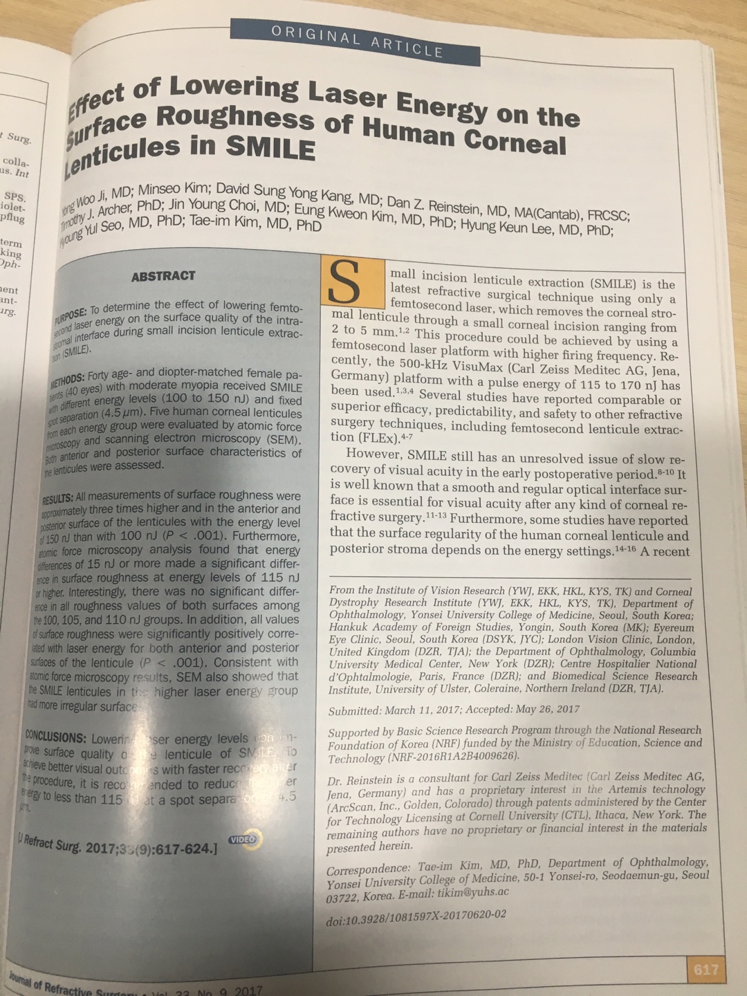
ORIGINAL ARTICLE
Effect of Lowering Laser Energy on the Surface Roughness of Human Corneal Lenticules in SMILE
ABSTRACT
Figure 2. Representative images of both surfaces of human lenticules by scanning electron microscopy. (A) The lenticule obtained from small incision lenticule extraction (SMILE) using conventional femtosecond laser energy (140 and 150 nJ) with a spot separation of 4.5 μm had a more irregular surface than that from SMILE using low energy (100 and 110 nJ) with the same spot separation on both the anterior and posterior surfaces. The posterior surface of the SMILE lenticule was rougher than the anterior surface at each energy level (x 300). (B) The cavitation bubbles were observed at the periphery of both surfaces of the lenticules from SMILE using laser energy of more than 120 nJ (x 300). to each other; some interface irregularity is inevitable and consequently affects visual outcomes. Our results revealed the anterior and posterior surfaces of the lenticule were significantly smoother when using lower energies. Moreover, the energy level had a highly positive correlation with the surface roughness of the anterior surface of the lenticule and posterior surface of the lenticule obtained from the SMILE procedure. Likewise, atomic force microscopy analyses of donor corneas undergoing preparation for endothelial keratoplasty showed that when lower energy lasers were used to dissect the donor tissue, the surface of the posterior stroma was more regular compared to when higher energy lasers were used.16 Also, it has been demonstrated that the surface regularity index decreased as laser energy increased during FLEx.14 Previous studies to evaluate the surface characteristics of FLEx or SMILE lenticules used only subjective criteria.12,14,15,19 Among them, some authors noted the smooth lenticule surfaces and the absence of surface irregularities by using environmental scanning electron microscopy for subjectively evaluating the anterior and posterior surfaces of SMILE lenticules.15 They used environmental scanning electron microscopy to eliminate artifacts due to tissue preparation; however, environmental scanning electron microscopy images are likely to look flat and smooth. Therefore, we used scanning electron microscopy for morphologic evaluation and objectively analyzed the surface roughness using atomic force microscopy. In contrast to the above study, our microscopic findings showed that the posterior surface of the lenticule was much rougher than the anterior surface of the lenticule. These are consistent with the scanning electron microscopy findings of other studies.12,19 Copyright © SLACK Incorporated
Lowering Laser Energy in SMILE/Ji et al
Energy Level A. Anterior surface A. Posterior surface B. Anterior surface B. Posterior surface 100 nJ SEM Image (100 μm) SEM Image (100 μm) 110 nJ SEM Image (100 μm) SEM Image (100 μm) 115 nJ SEM Image (100 μm) 140 nJ SEM Image (100 μm) SEM Image (100 μm) SEM Image (100 μm) 150 nJ SEM Image (100 μm) SEM Image (100 μm) SEM Image (100 μm)
Ji YW, Kim M, Kang DSY, Reinstein DZ, Archer TJ, Choi JY, Kim EK, Lee HK, Seo KY, Kim TI.
Abstract
PURPOSE:
To determine the effect of lowering femtosecond laser energy on the surface quality of the intrastromal interface during small incision lenticule extraction (SMILE).
METHODS:
Forty age- and diopter-matched female patients (40 eyes) with moderate myopia received SMILE with different energy levels (100 to 150 nJ) and fixed spot separation (4.5 μm). Five human corneal lenticules from each energy group were evaluated by atomic force microscopy and scanning electron microscopy (SEM). Both anterior and posterior surface characteristics of the lenticules were assessed.
RESULTS:
All measurements of surface roughness were approximately three times higher and in the anterior and posterior surface of the lenticules with the energy level of 150 nJ than with 100 nJ (P < .001). Furthermore, atomic force microscopy analysis found that energy differences of 15 nJ or more made a significant difference in surface roughness at energy levels of 115 nJ or higher. Interestingly, there was no significant difference in all roughness values of both surfaces among the 100, 105, and 110 nJ groups. In addition, all values of surface roughness were significantly positively correlated with laser energy for both anterior and posterior surfaces of the lenticule (P < .001). Consistent with atomic force microscopy results, SEM also showed that the SMILE lenticules in the higher laser energy group had more irregular surfaces.
CONCLUSIONS:
Lowering laser energy levels can improve surface quality of the lenticule of SMILE. To achieve better visual outcomes with faster recovery after the procedure, it is recommended to reduce the laser energy to less than 115 nJ at a spot separation of 4.5 μm.


