
Order of Examination
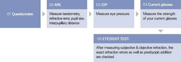
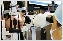
Cataract Examination Process
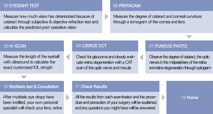

Eyesight Test
-

Questionnaire
Filling out our questionnaire is the most basic step of it. It allows us to find out your previous medical history and your family medical history. Also it enables us to know your exact condition and what vision problems you are experiencing.
-
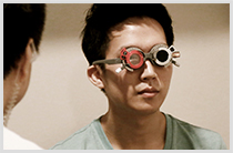
Eyeglass Test
Inspecting the exact strength of the eye glasses you are currently wearing helps us to measure your myopia, astigmatism, and hyperopia.
-

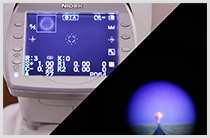
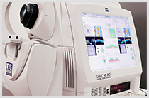
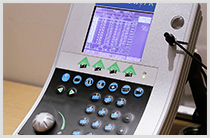
Automatic Refractive Power Test
Your exact refraction error is measured by used the most advanced optical equipment.
-

Manifest Refraction Test
Your maximum correct vision with glasses can be predicted with visual acuity test. The calculated maximum corrected vision is theoretically the maximum post-operative vision you will have.
-

Cycloplegic Refraction Test
Cycloplegic eye drops are used to temporarily paralyze the ciliary muscles that control focusing to determine your exact refractive error.
-
Subordinate Cycloplegic Refraction Test
After 3~5 days of the cycloplegic refraction test, when the medicinal effects have disappeared, another vision test is done. This is to calculate the change to more accurately measure myopia and astigmatism.
-
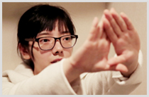
Dominant Eye Test
This is to find out which one of your eyes is the dominant one.
Cornea Examination
-

Corneal Stiffness Test
This test measures the corneal biomechanics (corneal stiffness – functional response to maintain original shape when under pressure). It is to prevent any side effects such as keratoconus after LASIK or LASEK surgery. All our patients are shown a comparison of their corneal stiffness before and after surgery.
-

Eyeglass Test
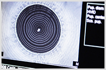
The corneal pattern/model and oblatenes, keratoconus and pupil size can be measured by examining the corneal curvature range and refraction range.
-
Automatic Refractive Power Test
Your exact refraction error is measured by used the most advanced optical equipment.
-
Manifest Refraction Test
Your maximum correct vision with glasses can be predicted with visual acuity test. The calculated maximum corrected vision is theoretically the maximum post-operative vision you will have.
-
Cycloplegic Refraction Test
Cycloplegic eye drops are used to temporarily paralyze the ciliary muscles that control focusing to determine your exact refractive error.
-

Subordinate Cycloplegic Refraction Test
Since it’s the most essential test for eyes condition, ‘doctor’ looks carefully at the cornea shape and whether or not these are going well.
Retina Examination
-

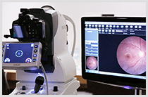
Optic Nerve & Retina Test
An optic nerve and retina CT scan diagnoses any retina diseases such as retina disorders, glaucoma and optic nerve damage
-
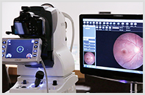
Fundus examination Test
The overall condition of the optic nerves and retina is reviewed to diagnose any retina degeneration, diseases from previous injuries and also the degree of retina damage from diabetes and high blood pressure.
-
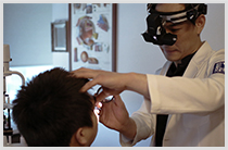
Retina Test
After enlarging the pupil, the optical nerves are examines with a special microscope. This test checks for any abnormality in the optical nerves or central retina area, glaucoma, injury related diseases, retina damage from diabetes or high blood pressure, and intraocular tumors.
Keratoconus Test
-

Automatic Refractive Power Test
Your exact refraction error is measured by used the most advanced optical equipment.
-

Manifest Refraction Test
Your maximum correct vision with glasses can be predicted with visual acuity test. The calculated maximum corrected vision is theoretically the maximum post-operative vision you will have.
-
Corneal Curvature Test
The corneal pattern and oblateness, keratoconus and pupil size can be measured by examing the corneal curvature range and refraction range.
-
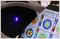
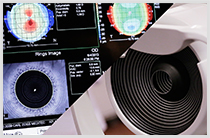
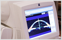
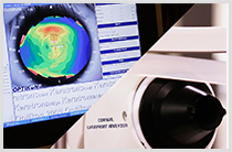
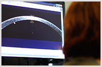
Corneal Topography Test
With a slit CT scan, the topography, curvature, thickness, shape and symmetry of the cornea are measured. This test enables a true customized surgery for each individual. Also, this test can detect irregular astigmatism, cataract, and glaucoma as well as measure the anterior and posterior corneal shape, cornea thickness, and front depth.
-

Corneal MRI Test
By displaying the exact condition of your cornea, iris, and lens in three dimensions, the corneal topography can be accurately measured. A simulation of the corneal flap thickness before and after surgery can be used to predict and prevent any side effects that could occur.
Glaucoma Test
-
Eye Pressure Test
The eye pressure is measure without actual contact. This allows for early diagnosis of glaucoma or to comprehend the exact condition of glaucoma and optic nerves.
-
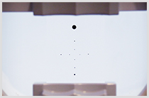
Comprehensive Visual Field Test MATRIX
This essential exam diagnoses glaucoma and any retina abnormality by checking light reaction and glaucomatous nerve damage.
-
Fundus examination Test
The overall condition of the optic nerves and retina is reviewed to diagnose any retina degeneration, diseases from previous injuries and also the degree of retina damage from diabetes and high blood pressure.
-
Comprehensive Visual Field Test HUMPHREY
If the results from the MATRIX test were negative, this test allows for a more thorough/detailed examination to check for not only glaucoma but also for retina abnormality and macular degeneration.
-
Optic Nerve & Retina Test
An optic nerve and retina CT scan diagnoses any retina diseases such as retina disorders, glaucoma and optic nerve damage
-
Cornea MRI Test
By displaying the exact condition of your cornea, iris, and lens in three dimensions, the corneal topography can be accurately measured. A simulation of the corneal flap thickness before and after surgery can be used to predict and prevent any side effects that could occur.
Night Blurred Vision Test
-
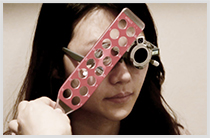
Night Pupil Size Test
People with excessive myopia experience night blurred vision mostly because the sizes of their pupils are much larger at night. So by measuring the exact size of your pupils in a dark surrounding, the possibility of post operative side effects can be predicted. Also with these measurements, your surgeon can decide on the size of the corneal flap
-
Automatic Pupil Size Test
This test measures the corneal pattern and oblateness, keratoconus and pupil size by examining the corneal curvature range and refraction range.
-
WaveFront Test
Using the Tscheming method that directly observes the WaveFront aberration, this test will analyze the anterior and posterior cornea as well as HOA, another cause that hinders vision beside myopia and astigmatism.
-

Automatic Refractive Power Test
Your exact refraction error is measured by used the most advanced optical equipment.
Dry Eye Test
-
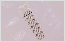
Tear Production Test
The Schirmer Test measures your tear production to predict the temporary dry eye condition you might experience after surgery.
-
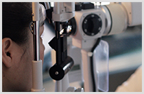
Slip Lamp Test
The binocular slip-lamp examination provided a stereoscopic magnified view of the eye structures in detail, enabling your specialist to diagnose various eye conditions.
-
Tear Breakup Time
Fluorescein is instilled into your tear film and the time that elapses between the last blink and the appearance of the first dry spot in the tear film is recorded. This clinical test is used to assess for evaporative dry eye disease.
Cataract Test
-
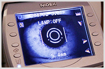
A Scan Test
Ultrasound if used to determine the exact condition of your eyes. Information needed for cataract surgery or IOL surgery such as the length of your eyeball, frontier depth, any existence of foreign substances, and your retina are measure and evaluated.
-

Manifest Refraction Test
Your maximum correct vision with glasses can be predicted with visual acuity test. The calculated maximum corrected vision is theoretically the maximum post-operative vision you will have.
-

Slip Lamp Test
The binocular slip-lamp examination provided a stereoscopic magnified view of the eye structures in detail, enabling your specialist to diagnose various eye conditions.
-

Automatic Refractive Power Test
Your exact refraction error is measured by used the most advanced optical equipment.
-

Eye Pressure Test
The eye pressure is measure without actual contact. This allows for early diagnosis of glaucoma or to comprehend the exact condition of glaucoma and optic nerves.
-

Corneal Topography Test
With a slit CT scan, the topography, curvature, thickness, shape and symmetry of the cornea are measured. This test enables a true customized surgery for each individual. Also, this test can detect irregular astigmatism, cataract, and glaucoma as well as measure the anterior and posterior corneal shape, cornea thickness, and front depth.
-

fundus examination
The overall condition of the optic nerves and retina is reviewed to diagnose any retina degeneration, diseases from previous injuries and also the degree of retina damage from diabetes and high blood pressure.
Consultation
-
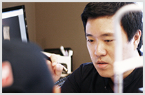
Analyze Test Results
The exact condition of your eyes can be assessed with the results from the various comprehensive tests, so the most suitable surgery can be chosen for your eyes. Surgery method, recovery time, and any precautions before and after surgery will be informed, and any question and inquiries you might have will be answered.
-
Customized Surgery Consultation
A one-on-one consultation with your own private specialist will take place. You and your specialist will examine the results together to choose the right surgery method for you. The specialist that you consulted with will be your surgeon who will perform your surgery.